

Have you ever looked at a science diagram, maybe a picture of a cell dividing, and wished it made more sense? You're not alone. It can feel a bit like trying to read a map without street names. But when you get a mitosis model labeled just right, suddenly the whole process of how cells make copies of themselves clicks into place. This is a really important idea in biology, you know, because it explains how we grow and how our bodies fix themselves. So, we're going to take a closer look at these models and see how they help us figure out the amazing story of cell division.
Cell division, or mitosis, is a truly central part of how life works. It’s how one cell becomes two, and then four, and so on. My text tells us that mitosis is a key part of the cell cycle, a series of stages that helps cells divide and pass on their genetic information. It's a very precise operation, actually, ensuring each new cell gets a perfect set of instructions.
Learning about mitosis with a clear, mitosis model labeled helps you see exactly what's happening at each step. It makes the invisible visible, giving you a picture to connect with the words. We’ll go through the different parts of this process, seeing what each stage looks like, and why getting these labels right makes all the difference for your understanding.
Table of Contents
- Understanding the Cell Cycle First
- The Purpose of Mitosis
- The Four Main Stages of Mitosis on Your Model
- How the Cell Cycle is Controlled
- When Things Go Wrong: Cell Division and Health
- Making the Most of Your Labeled Model
- Frequently Asked Questions About Mitosis Models
Understanding the Cell Cycle First
Before we jump into mitosis itself, it's good to remember that mitosis is just one part of a bigger cycle. My text mentions that in eukaryotic cells, there are clear phases of the cell cycle. This whole cycle is how a cell grows, gets ready to divide, and then actually divides. It’s pretty much the life story of a cell.
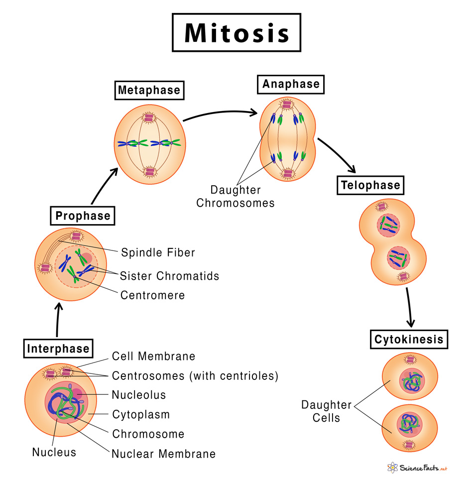
A cell spends most of its time in a phase called interphase. This isn't part of mitosis, but it's where the cell does a lot of important work to prepare. Interphase has three main parts, you know. First, there's the G1 phase, where the cell grows and takes in nutrients. This is like the cell just getting bigger and stronger, in a way.
Then, we have the S phase. This is a very important part because, as my text says, the cell's DNA is replicated here. Think of it like making a perfect copy of all the cell's instruction manuals. After that, there's the G2 phase, where the cell prepares for division. It's almost like a final check before the big event. All of this happens before mitosis even begins, which is quite interesting.
The Purpose of Mitosis
So, why do cells go through all this trouble? The main goal of mitosis, according to my text, is to produce daughter cells that are genetically identical to their mothers. This means the new cells have the exact same number of chromosomes, without any extra or missing ones. It's a very precise process, really, ensuring genetic information is passed on perfectly.
This is super important for many reasons. For one, it’s how we grow from a single cell into a complex organism. It's also how our bodies replace old or damaged cells. Think about a cut on your skin; new skin cells are made through mitosis to heal it. So, it's about growth, repair, and just keeping things going.
My text also briefly mentions meiosis, but that’s a different kind of cell division, used for a different purpose, you know, for reproduction. Mitosis is all about making exact copies for growth and maintenance. It's like having a photocopy machine that always produces perfect duplicates.
The Four Main Stages of Mitosis on Your Model
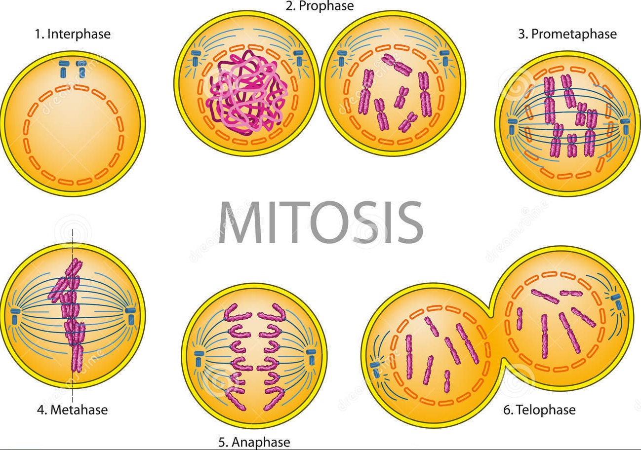
My text tells us that mitosis involves a series of stages: prophase, metaphase, anaphase, and telophase. Some textbooks, it says, mention five stages because they separate prophase into an early phase (just called prophase) and a later one (sometimes called prometaphase). For our mitosis model labeled, we'll focus on the main four, which are typically what you see represented.
When you look at a labeled diagram of mitosis, each stage shows distinct changes in the cell. Knowing what to look for at each step helps you understand the whole picture. It’s like watching a play, where each act has its own set and actions. Let's break down what you should expect to see.
Prophase: The Start of the Show
Prophase is where things really start to get organized inside the cell. On your mitosis model labeled, you'll see the genetic material, which was all spread out during interphase, begin to condense. It coils up, becoming shorter and thicker, so it's easier to move around. These condensed structures are what we call chromosomes.
You might also notice that the nuclear envelope, which is like the membrane around the cell's genetic material, starts to break down. This prepares the way for the chromosomes to be moved. Also, tiny structures called spindle fibers begin to form. They're sort of like ropes that will help pull the chromosomes apart later. It’s a very busy time for the cell, getting everything ready.
Metaphase: Lining Up for Division
Next up is metaphase, a very distinct stage on any mitosis model labeled. What you'll notice immediately here is that the chromosomes line up perfectly in the middle of the cell. They form a straight line, almost like soldiers standing at attention. This is often called the metaphase plate.
The spindle fibers, which started forming in prophase, attach to each chromosome at a specific spot. This precise alignment is really important, you know, because it makes sure that when the cell divides, each new cell gets an exact copy of every chromosome. It’s a moment of great order before the separation begins.
Anaphase: Pulling Apart
Anaphase is a dramatic stage to see on your mitosis model labeled. Here, the sister chromatids, which are the two identical halves of each chromosome, finally separate. The spindle fibers, which are attached to them, start to shorten and pull these separated chromatids to opposite ends of the cell. It's a very active process, with the cell stretching out a bit.
Each chromatid, once separated, is now considered a full chromosome on its own. So, you have one complete set of chromosomes moving towards one pole of the cell and another identical set moving towards the opposite pole. This ensures that the genetic material is evenly split between the two future daughter cells. It’s a pretty quick phase, actually, but very important.
Telophase: Two New Cells in the Making
Telophase is the final main stage of mitosis. On your mitosis model labeled, you'll see that the chromosomes have arrived at the opposite poles of the cell. They start to uncoil and become less condensed again. New nuclear envelopes begin to form around each set of chromosomes, creating two distinct nuclei within the single cell. This is where the cell really starts to look like it's preparing to become two separate entities.
At the same time, the spindle fibers disappear. You might also notice that the cell itself starts to pinch in the middle. This pinching is part of cytokinesis, which is the actual physical division of the cell's cytoplasm. While technically separate from mitosis, cytokinesis usually happens right after or during telophase, completing the process of forming two new, independent cells. It's the grand finale, so to speak.
How the Cell Cycle is Controlled
The cell cycle, including mitosis, doesn't just happen randomly. It's very carefully controlled by the cell itself. My text points out that we’ll look at how the cell cycle is regulated. There are specific checkpoints in the cycle, you know, like quality control stations, that make sure everything is going as it should before the cell moves to the next stage.
These checkpoints look for things like whether the DNA has been copied correctly, or if the chromosomes are properly lined up. If there are any problems, the cell can pause the cycle to fix them. This control system is absolutely vital for keeping our bodies healthy and functioning properly. It’s a bit like a very smart internal clock with built-in safety mechanisms.
When Things Go Wrong: Cell Division and Health
What happens if this careful control system breaks down? My text says we’ll examine how disruptions can lead to cancer. When cells lose their ability to regulate their division, they can start to divide uncontrollably. This uncontrolled growth is a hallmark of cancer. It's a very serious consequence when the normal rules of cell division are ignored.
Understanding the normal process of mitosis and how it's controlled is therefore a very important step in understanding diseases like cancer. Scientists study these processes to find ways to stop or slow down uncontrolled cell growth. So, your mitosis model labeled isn't just about basic biology; it connects to real-world health issues, which is quite powerful.
Making the Most of Your Labeled Model
To truly get the most out of your mitosis model labeled, take your time with it. Don't just glance at it. Trace the path of the chromosomes through each stage. See how they change from being spread out to condensed, then lined up, pulled apart, and finally forming two new groups. This active engagement helps to solidify your understanding.
You could even try to draw each stage yourself, labeling the key parts. This is a very effective way to learn, as it forces you to pay attention to the details. Compare your drawings to your model and to other resources. For example, Khan Academy offers free resources on the cell cycle and its role in growth, development, and reproduction, which can be a great external resource to cross-reference your model with. You can learn more there.
Also, try to explain each stage to someone else, or even just to yourself. If you can explain it clearly, it means you really understand it. A good mitosis model labeled is a fantastic tool for this kind of active learning, making a complex topic much more approachable. It’s a pretty neat way to get a solid grasp on cell division.
Frequently Asked Questions About Mitosis Models
What's the main difference between mitosis and meiosis on a model?
Basically, a mitosis model shows one cell making two identical copies. A meiosis model, on the other hand, would show a cell making four different cells, which have half the usual number of chromosomes. My text tells us meiosis is only used for a specific purpose, you know, unlike mitosis which is for growth and repair.
Why do some mitosis model labeled diagrams show five prophase stages?
My text explains that some textbooks mention five phases because they separate prophase into an early phase, just called prophase, and a later one. This later phase is often called prometaphase. It just adds a bit more detail to the very start of the division process, showing the nuclear envelope completely breaking down and spindle fibers fully attaching.
How can I tell if a cell on a model is in interphase or mitosis?
You can usually tell by looking at the genetic material. In interphase, especially the S phase, the DNA is replicated, but it's still spread out and not visible as distinct chromosomes. During mitosis, however, the chromosomes become very condensed and visible, especially as they line up and separate. So, if you see clear, rod-like structures, it's likely a mitotic stage.

Detail Author:
- Name : Guiseppe Kuhic
- Username : vada.denesik
- Email : dennis12@cole.com
- Birthdate : 1982-10-03
- Address : 4453 Heaney Ramp Apt. 281 Runtefort, VA 72837-7745
- Phone : 415-988-0266
- Company : Bosco-Halvorson
- Job : Municipal Fire Fighting Supervisor
- Bio : Consequuntur in porro dolorem aut quas sed minus. Aperiam tempore fugit voluptatem. Optio placeat et sit itaque ipsum ut ipsa eaque. Quo rerum voluptas harum quam non odit quasi.
Socials
twitter:
- url : https://twitter.com/ned_official
- username : ned_official
- bio : Harum ea voluptates atque est. Consequatur ut debitis ut maiores officiis quo.
- followers : 6328
- following : 448
facebook:
- url : https://facebook.com/nschroeder
- username : nschroeder
- bio : Deserunt et et ipsa quibusdam odio.
- followers : 258
- following : 1204
tiktok:
- url : https://tiktok.com/@nschroeder
- username : nschroeder
- bio : Nihil aspernatur nihil saepe.
- followers : 5074
- following : 1750


 Bonus
Bonus